Time-of-Flight Secondary Ion Mass Spectrometry (ToF-SIMS)

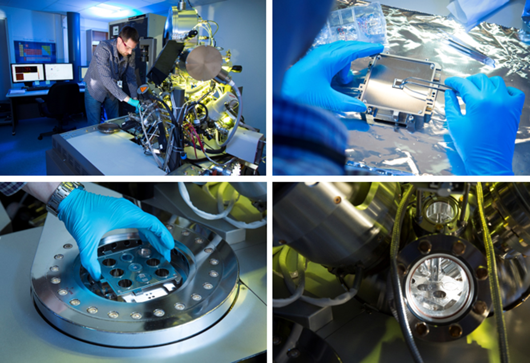
Our Time-of-Flight Secondary Ion Mass Spectrometry (ToF-SIMS) instrument (Trift V nano TOF from Physical Electronics) allows us to detect small amounts (down to 1 ppm) of any element or molecule on a surface. The lateral resolution is below 1 µm, so chemical maps can be made, showing where a certain element or molecule is present. The instrument has a built-in Focused Ion Beam (FIB) which can cut into the sample surface and reveal the three-dimensional structure of the material.
ToF-SIMS spectra are generated using a pulsed ion gun which shoots a focused beam of primary ions onto the sample surface. This causes a cascade of collisions in which some atoms are kicked out from the surface. A small fraction of the atoms gain or loose an electron in the process. These secondary ions are accelerated and then travel through the ToF analyzer with different velocities, depending on their mass to charge ratio.
For each primary ion pulse, a full mass spectrum is obtained by measuring the arrival times of the secondary ions at the detector. The ion masses are determined with high precision after calibrating with know ions.
By scanning the primary beam in a raster pattern across the surface, we get mass spectra from every pixel in the imaged area. By using a separate sputtering gun to remove some of the material between each image, we can obtain three dimensional information from the sample.
These data can be displayed in three ways (the three modes of SIMS):
- Mass spectra (showing the number of counted ions of a given mass)
- Ion images (showing where any selected element or isotope is present in the imaged area)
- Depth profiles (showing the count rate for any isotope as a function of time time at increasing sputtering depth).
The additional FIB gun can make tilted cross sections several micrometers into the surface. These freshly cut surfaces can then be analyzed (see image below). By making a new FIB cut and re-analyzing, the full 3D structure of the material can be determined.
Relevant links
Infrastructure networks:
Projects: