The new operating method is based on an ultrasound-based navigational system developed through an interdisciplinary collaboration over many years between technologists and physicians at SINTEF, NTNU, St. Olav’s Hospital and the company, Mison.
The system produces a “road map” or, more precisely, a “body map” comprising 3D MR pictures taken of the patient prior to the operation. These pictures are saved on a computer. The objective of the system is to connect these pictures with the live pictures taken when the patient is on the operating table.
The three-dimensional pictures appear on a screen in the operating theatre – just like GPS navigation in a car. The system traces the position of the surgeon’s operating instruments and plots them on the ”body map” in the same way that a car’s position is plotted on a map in the car.
| 
|
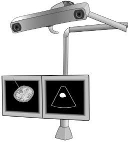
To track the position of the ultrasound (US) probe and surgical instruments, the navigation system radiates infra-red (IR) light from an arm-mounted camera. The navigation tools are equipped with four reflecting spheres, which are detected by the system. The position of the instrument is indicated in image slices extracted from 3D MRI or 3D US volumes of the patient (left monitor). Conventional 2D US is shown on the right monitor. (ill. OM Rygh) | |
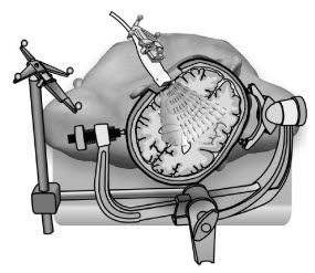
As the surgeon operates, removal of the tumour mass and loss of spinal fluid leads to movement in the brain and, as a consequence, imprecise navigation. The map the surgeons are using to guide them must then be updated with new pictures. These new pictures are produced with the aid of an ultrasound probe, which is used during the entire operation. The probe sends sound signals like an echo sounder, and provides pictures that show the brain’s landscape as the operation is taking place. The new pictures enable the surgeon to navigate the instruments with great precision. As a result, the chances of damage to normal brain tissue is reduced, it is easier to locate the tumour and to remove more of it.

