|
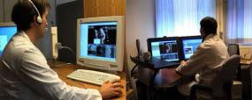
X-ray specialists in different hospitals can interpret the X-ray images together, just as if they had been sitting in the same room. |
Norwegian and American IT specialists have built one of the first telecommunications of its kind in the world. The system gives radiologists the opportunity to interpret three-dimensional x-ray images together - each of them working in his own hospital - via a broadband network.
In a pilot project, the new advance was used to link the x-ray departments at Molde Hospital and St. Olav’s Hospital in Trondheim.
Saves journeys and overnight stays in hospital
So far, the system has been tested on five patients from Romsdal, saving them long trips to the hospital in Trondheim for post-op check-ups. Now they can stay in Molde instead and be checked up there by remote control, because the x-ray specialists at St. Olav’s “travel” to the patients via the Internet. This solution also saves Norwegian hospitals expensive overnight admissions.
Cancer patients next?
The system is currently being used to follow up patients who have been operated for abdominal aortic aneurysm. This is a relatively small group of patients. Most of them are elderly and they are very glad to have the long trip for their post-op check-ups shortened.
The next step could see the system making savings on a much larger group of hospital admissions. According to SINTEF scientist Jon Harald Kaspersen, who has been involved in developing the new system, this is because it would be very suitable for similar collaboration between large and small hospitals when radiation treatment of cancer patients is being planned.
Presentation at world congress
SINTEF has developed the system in collaboration with the American IT company Kitware. Recently the partners presented the system at the world congress of radiologists in Chicago, which brought together some 60,000 delegates, 200 of whom attended the session at which the Norwegian-American system was demonstrated via a transatlantic link.
Checking “internal repairs”
The Romsdal patients who are being followed up by cyberspace have all been operated for abdominal aortic aneurysm at St. Olav’s Hospital. These patients have all had their aorta repaired from the inside.
In operations of this sort, the surgeons introduce their instruments through the blood vessel from the groin. From inside the vessel, they install a “stent”, a sort of scaffolding, on the damaged part of the vessel wall. This type of operation has less serious effects on the body than traditional open surgery.
Specialists travel - via the Internet
The patients would normally need to travel back to Trondheim twice a year for post-op 3D x-ray check-ups of their “scaffolding”. Now, the x-rays can be made in Molde instead.
With the aid of the technology from SINTEF and Kitware, the radiologists from the two hospitals can study the three-dimensional images together via broadband link. Each of them can control the field of view from his own hospital, zooming in and examining different aspects, looking at the same part from his own end of the link and interpreting the images in conjunction.
Efficient training method
“It is just like sitting in the same room and looking at the x-ray plates together”, says Jon Harald Kaspersen. According to the SINTEF scientist, the radiologists involved in the project say that the system provides a very efficient method of training at the local hospital. “In the long term, the system will enable the doctors in Molde to follow up these patients on their own”.
The system is a result of close collaboration between technologists and hospital doctors in Trondheim. “This solution could never have come to fruition without such cooperation”, says Kaspersen. The pilot project in mid-Norway is being financed by the Research Council of Norway, as part of the HØYCOM Project.
Contact:
{DynamicContent:Ansatt link}
